
A volumetric analysis of the brain is very straightforward. We know that certain things such as aging, brain injury, stroke and dementia can cause a reduction in the size of certain parts of the brain.
Page Contents
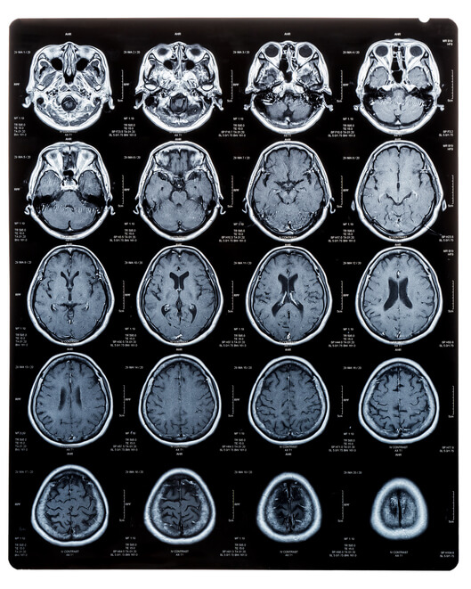
VOLUMETRIC BRAIN MRI
Medical science has over the past 25 years been able to perform numerous MRI studies of the brain on thousands of individuals to develop what is called a normative data base in order to determine what the average or normal brain volume is for various ages and both genders over time. When this is known, it permits brain scientists and doctors to then measure brain volume in patients with medical conditions that are known to cause volume loss in the brain, and compare those patient’s MRI scans to the normative data base to see whether volume loss has occurred.
If volume loss has occurred, then a patient’s doctor can determine what the cause is. One well known cause of volume loss is brain injury. Brain injury causes volume loss because damaged axons that ultimately die off will shrink in size. When a group of such cells are damaged in an area of the brain susceptible to brain trauma, collectively they can, over time, show a measurable loss in volume of that particular spot in the brain. We see the same phenomena when a person has a stroke, and an entire area served by a blood vessel dies off due to lack of blood and oxygen. As a result of the dying off of brain cells, the area affected shrinks and can be clearly measured against normative data bases.

With Brain injuries, we see the same effect. Because certain parts of the brain are more prone to damage when a brain injury occurs, these are the areas that we typically see volume loss occur. If a person has asymmetric, or uneven volume loss in these areas versus a more globally diffuse pattern of volume loss seen in dementia, we know it is more consistent with trauma causing that damage. For example, gray matter and white matter have different densities, so when trauma occurs, we can often see damage at the gray/white matter junctions due to those differences. If volume loss shows up 18 months after the head trauma in these areas, radiologist and doctors will agree that this is more consistent with brain trauma than any other disease process.
At Brain Injury Law of Seattle, this is one more tool we use to help objectively prove brain injury in addition to the other various modalities we use. For more details contact us today!


